All posts by derm

Lichen planus

Lichen planus is an inflammatory disease that manifests itself in pink-fillet papules on the skin. The lesions are often located on the straight surface of the forearms but may occur in any other location.

Lichen planus can also occur within the oral mucosa and genital mucosa (white linear structures, redness or erosions). Changes in the genitals may cause severe itching. The lichen planus of the scalp (lichen planopilaris) is associated with inflammation around the hair follicles and may cause permanent alopecia if adequate treatment is not started early enough. A subtype of lichen planopiliaris is frontal fibrosing alopecia, which consists of gradual shifting of the hairline towards the back. This form of hair loss can have a very similar clinical manifestation to androgenic alopecia. The diagnosis is determined by the trichoscopic examination and in some cases, a histological examination of the scalp (biopsy) may be required.

In our office, the treatment of lichen planus is led by Prof. Małgorzata Olszewska. Prof. Lidia Rudnicka and Dr. Adriana Rakowska treat lichen planus when there is involvement within the scalp.

Fungal infections

Mycosis is caused by infection with pathogenic fungi and may have various symptoms.

Dermatologists deal with the diagnosis and treatment of diseases such as:

  • athlete’s foot
  • onychomycosis
  • athlete’s hand
  • inflammation of the groin
  • mycosis of smooth skin
  • tinea scalp
  • athlete’s foot
  • dandruff versatile.

Diagnosis of tinea is based on a clinical examination and medication trial, which in many cases is sufficient. Additional tests include mycological examination (the result is obtained after several weeks) and sometimes a clipping (biopsy) for histopathological examination. We perform trichoscopy in cases of suspected mycosis of the scalp.

Trichoscopy

Trichoscopy is an innovative method of diagnosing hair and scalp diseases. The method is based on dermoscopy. In trichoscopy hair and scalp structures can be visualized at multiple magnification levels and a special light source allows for partial analysis of the structures under the skin. Trichoscopy is performed only by doctors with extensive dermatological experience and deep medical knowledge, which allows for proper interpretation of trichoscopic images. The basic trichoscopy can identify most of the important irregularities. The result of such an examination is taken into account by the physician when making treatment decisions. The so-called ‘full trichoscopy’ involves quantitative analysis of abnormalities in some hair diseases. This test is useful before starting treatment and to monitor the effectiveness of treatment in diseases such as androgenic alopecia or telogen effluvium.

Prof. Lidia Rudnicka was the first person to describe trichoscopy and developed criteria for recognizing many diseases using this method. Currently, trichoscopy is used by dermatologists around the world.

Prof. Lidia Rudnicka is a co-founder and the first president of the International Trichoscopy Society. Warsaw was chosen to be the city of the first world trichoscopic congress.

Dermoscopy

Dermoscopy (dermatoscopy) is a painless, non-invasive microscopic examination of the skin, which does not require a skin biopsy. It is intermediate examination that involves a normal clinical assessment and a histopathological examination of skin.

Dermoscopy offers the possibility of viewing the skin in a ten-to-ten-fold magnification and in lighting, which allows the dermatologist to see the deeper structures of the epidermis.

Dermoscopy is commonly used to assess pigmented nevi, commonly referred to as moles. This method increases the ability to recognize melanoma at a very early stage of development, where this cancer can be easily cured. In addition, dermoscopy makes it easier to distinguish melanoma from other harmless lesions on the skin.

Computer dermoscopy (videodermoscopy) is becoming more and more widely used in dermatology. This method has the additional advantage of showing the examined part of the skin on the computer screen and allows for saving this image in electronic form. This is particularly important in the study of patients who have a lot of pigmented nevi. During control examinations, carried out once a year, it is possible to compare the appearance of each mole to its appearance in previous years. If the birthmark grows or changes, the dermatologist may decide to remove it. Videodermoscopy is worth doing in the same dermatology office so that it is possible to compare the signs to those that are already in the database. It is also worth asking the doctor whether he is saving marks in the database. The visualization of a mark on a computer screen is not tantamount to the fact that it has been cataloged and saved.

Dermoscopy can predict whether a small nevi may develop into melanoma with up to 95% accuracy. There may always be lesions that do not fulfil all melanoma criteria in dermoscopy, but because of some melanoma characteristics, they raise concern . Such lesions are always preventively removed.

The price of dermoscopic examination for pigmented nevi depends on the number of lesions with a range between 250 to 600 PLN.

Recently, dermoscopy has become a diagnostic method widely used also in other areas of dermatology. It gives you not only the ability to study pigmented birthmarks and early detection of melanoma, but also to watch almost all other skin lesions. This method can be used for the study of capillaroscopy (examination of blood vessels of the nail shaft) in people with suspected Raynaud’s disease, scleroderma or dermatomyositis. Dermoscopy can also be used to view the hair follicles, and in the video-onodoscopic examination, hair diameter can be measured, which is used in some cases of diagnostic problems related to hair diseases.

Diseases of nails

Nail disease can be an independent medical problem, not related to other symptoms. An example of such disease is onychomycosis.

Onychomycosis usually affects the nails of the feet, but it can also occur within the nails of the hands. Nails change color, become thickened and fragile. Treatment consists of general antifungal drugs. Changes disappear slowly, usually at the rate at which the nails grow, which is usually 1 mm per month.

Similar symptoms can occur in nail psoriasis. In these cases, they often accompany psoriatic lesions on the skin. Usually, general medications in tablets or injections are used in the treatment.

Another common nail disease is lichen planus. In this disease the most common symptom is the development of longitudinal grooves within the nail plates. Other unique symptoms of lichen planus may also occur.

Dr. Marta Sar-Pomian specializes in nail diseases and surgery. She also performs diagnostic biopsies of nail changes and nail dermoscopy.

Vitiligo

In vitiligo there is lack of pigment, known as ‘white spots’ (maculas). About 1-2% of the population suffers from vitiligo. Most often, the first white spots appear between 10 and 30 years old, but children can also suffer from vitiligo.

The cause of this disease is unknown. Genetic predisposition may play a role in development of vitiligo, but most often neither parents nor children of people with vitiligo have any symptoms of this disease.

As a result of the destruction of cells responsible for the production of melanin, white maculas are formed. Destruction of melanocytes, which produce the melanin, occurs immunologically. In vitiligo, the immune system mistakenly recognizes melanocytes in the skin as harmful to the body and gradually destroys them. In some people, the skin may rebuild melanocytes in those depigmented areas after proper treatment.

The manifestation of vitiligo depends on the severity of the disease. It may manifest itself only in the transient occurrence of one or more spots on the skin, while in extreme cases even discoloration of the whole skin may occur.

Most often white spots are formed on the face, neck, forearms, wrists and hands, around the armpits and genitals. Lesions can also occur within the scalp. Discoloration may also occur in the hair (white-gray strands), eyebrows and eyebrows.

Vitiligo begins with the appearance of small lesions of discolored skin, which are gradually enlarged. White spots are more sensitive to solar ultraviolet radiation and are easier to burn. But this does not mean that people suffering from vitiligo should completely give up contact with the sun. On the contrary, a small and careful exposure to the sun could support the treatment. Ultraviolet (UVA) lamps are even used to treat vitiligo.

Other methods of treatment of vitiligo include creams and ointments with immunomodulatory effects, and in some cases, appropriate oral medications are used. In any case, the clinical decision making for the treatment depends on a detailed analysis of the course of this disease, the severity of changes and the results of laboratory tests.

In our office, patients with vitiligo are primarily treated by Prof. Lidia Rudnicka and Dr. Małgorzata Łukomska.

Children

PEDIATRIC DERMATOLOGY

We also diagnose and treat skin diseases in children.
Diagnosis and treatment of diseases of young children are mainly dealt with by the Dr. Małgorzata Łukomska.
Dr. Małgorzata Łukomska has completed three specializations: pediatrics, dermatology and infectious diseases. She has experience in hospital and ambulatory treatment of skin diseases in children.
Investigations of pigmented nevi (moles) in children are performed by Dr. Justyna Sicińska, who specializes in the early detection of melanoma. The tests are performed using the videodermoscopy method. Videodermoscopy (videodermatoscopy) allows for the monitoring of pigmented birthmarks and the assessment, at the next examination, whether the birthmark has increased or changed.
Research on congenital hair diseases in children is carried out by Dr. Adriana Rakowska. The tests are performed using the trichoscopy method. Trichoscopy is a modern technique that does not require pulling hair for examination under a microscope.
Minor medical treatments for children are performed by Dr. Justyna Sicińska.

Aphthae (oral ulcers)

More than 2,400 years ago, Hippocrates described for the first time intensified mouth ulcers and called them “aphthae”, which means “to light” or “to kindle a fire” in Greek. This term referred to the ailments that accompany oral ulcers. Every third person has aphtha at least once in their lifetime. A single aphtha heals in a few days and leaves no trace. It may even occur unnoticed. Patients should see a dermatologist if they develop several aphtha. Some individuals may have ‘recurrent aphthosis’, where the healing of one aphtha on the mucous membrane leads to the emergence of another.

There may be many reasons of recurrent aphthosis, including mechanical injuries, stress, certain foods, infections and other diseases. Sometimes, despite precise examination, these causes can not be determined. The genetic factors and family predisposition play an important role in the onset of recurrent aphthosis. About 30% of people with recurrent aphthosis have a family member with the same disease. Children whose parents both have had aphthosis carry a 90% increased risk for its development.

The beginning of the disease usually occurs in childhood or adolescence. Usually, the first oral ulcers appear before the age of 30. Aphthae can result in small painful erosions or superficial ulcerations, usually up to 1 cm in diameter, which are yellowish or gray in coloration. These painful erosions or ulcerations are surrounded by an inflammatory rim. Typically, single aphtha heals in 7 to 10 days. If a single mucosal lesion persists longer, it may be a special form of aphthosis, but is probably not an aphtha.

In doubtful cases, other diseases should be excluded, such as erythema multiforme, lichen planus, chronic ulcerative stomatitis or pemphigus.

Prof. Małgorzata Olszewska, among others, deals with the treatment of aphthae and other mucosal diseases in our office.

Contact

Dermatology and Aesthetic Center

SPECJALIŚCI DERMATOLODZY S.C.

Grójecka Street 186/309
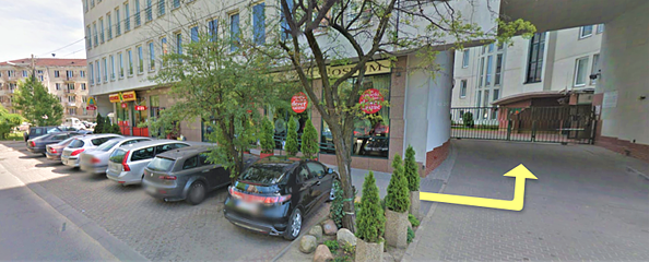
(Colosseum Building; entrance from Włodarzewska Street)
02-392 Warsaw

tel. (022) 824 22 00; 0-601-98-00-48 (text)
fax: (022) 824 22 00
e-mail: recepcja@dermatolodzy.com.pl


OPENING HOURS:

Monday-Friday 08:00-20:00

Saturdays 09:00-15:00

Wejście


SEND A MESSAGE:

CONSULTATIONS

We diagnose and treat all diseases of the skin, nails, hair and mucous membranes of the mouth and tongue. Each doctor has experience in establishing diagnoses and treatment plans for a full spectrum of skin diseases. Below are the specific interests of individual doctors:

  • Acne
  • Alopecia areata
  • Seborrheic dermatitis
  • Dandruff
  • Rosacea
  • Psoriasis
  • Purulent skin diseases
  • Aphtae, diseases of the oral mucosa
  • Allergic skin diseases
  • Skin diseases in children
  • Diseases of nails
  • Melanoma
  • Tinea capitis, onychomycosis, and other fungal infections
  • Consultations and procedures in aesthetic dermatology
  • Skin cancers
  • Herpes simplex
    Pemphigus, pemphigoid and other bullous diseases
    Systemic sclerosis (scleroderma) and morphea
  • Hair loss – differential diagnosis and treatment
  • Systemic lupus erythematosus
  • Nevi / moles, skin cancer prevention
  • Zoster